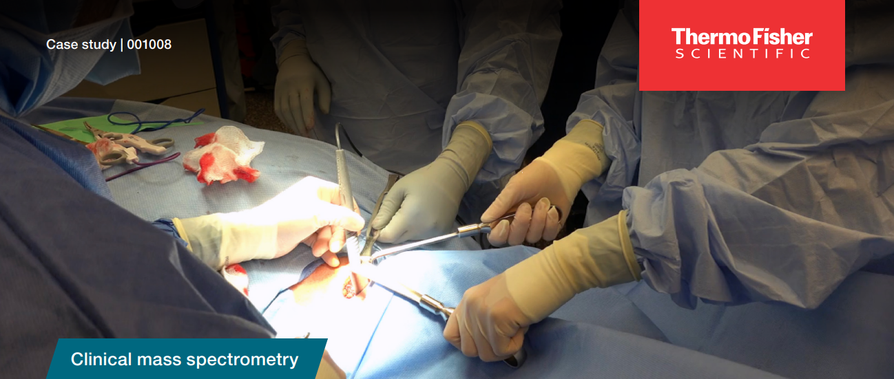
The MasSpec Pen in the hands of surgeons

In this application note, Thermo Fisher Scientific explores the potential benefits of mass spectrometers over microscopes in surgical histopathology. By utilizing spectra instead of traditional stains, surgeons and pathologists could achieve real-time identification of cancerous versus normal tissue, leading to reduced surgical time and enhanced patient care. The conventional method involves freezing, sectioning, and staining tissue samples, followed by microscopic analysis, a process taking 15–45 minutes per sample. However, this approach has limitations, including artifacts and complexities in certain tissues like pancreatic cancer. Mass spectrometry offers a promising alternative for faster and more accurate intraoperative tissue analysis.

